Lafayette has secured a $205,020 grant from the National Science Foundation (NSF) for a state-of-the-art desktop scanning electron microscope.
The microscope is expected to be used for analysis in many areas of research, including those related to engineering, chemistry, materials, geosciences, and biology.
Professors will use it in areas such as materials study, microfluidic and/or lab-on-chip systems, semiconductor devices, fossilized samples relevant to ancient ecology and climate, and biological and/or bioengineered systems.
The instrument will also be put to use in various core, laboratory-based, design, and/or elective courses in both chemical & biomolecular engineering and geology & environmental geosciences. Depending on the course, students will either have hands-on experience using the microscope, or see it used by instructors for demonstration purposes. Students working on research for honors thesis, independent study, and EXCEL Scholars projects will also have opportunities to use the instrument.
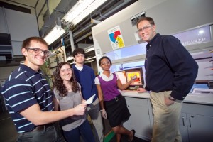
Joshua Levinson, right, assistant professors of chemical and biomolecular engineering, works with students in his Design Synthesis course.
Joshua Levinson and Chris Anderson, assistant professors of chemical and biomolecular engineering, and David Sunderlin, associate professor of geology and environmental geosciences, prepared the grant application.
“Until this award, scanning electron microscope (SEM)-based imaging was not available at the College, and this SEM will now provide the campus with a very useful tool for a wide array of active research areas,” Levinson says.
The Phenom ProX Desktop SEM, manufactured by NanoScience Instruments, Inc., has a magnification range of 80x–100,000x, with a resolution that is less than or equal to 17 nanometers. The equipment is expected to arrive later this semester and will be set up as soon as possible.
“Generally speaking, this system lends itself to the imaging and characterization of samples and sample surfaces at the sub-millimeter scale, which is critical to the thorough understanding of the dimensions, form, and function of the objects being studied,” Levinson says.
This compact system possesses a user-friendly interface that incorporates both a keyboard and a touch-screen and has quick loading-to-imaging times. Such features make it ideal for use at the College.
The microscope can be used to perform electron dispersive spectroscopy (EDS) for elemental identification. Associated software can be used for elemental mapping of sample surfaces using EDS. Other software that is incorporated into the system can be used to analyze the dimensions of fiber-based samples and to reconstruct three-dimensional surface roughness. Example applications of each include determining the composition of mineral particles, calculating accurate fiber- and pore-size measurements for biomimetic polymer matrices, and the sub-micron structure and mean roughness of etched surfaces, respectively.
The equipment will include a motorized tilt and rotation stage, a temperature-controlled stage for imaging moist/water-containing samples, and an array of sample holders designed to accommodate different types of samples.
